Month: March 2010

  • Collateral Damage by Mitochondria

    This month, I read a paper (HT: my ex-college roommate Eric) by a group from Beth Israel about systemic inflammatory response syndrome (SIRS) following serious injury. SIRS, which is more commonly understood/found as sepsis, happens when the entire body is on high “immune alert.” In the case of sepsis, this is usually due to an infection of some sort. While an immune response may be needed to control an internal infection, SIRS is dangerous because the immune system can cause a great deal of collateral damage, resulting in potentially organ failure and death.

    Whereas an infection has a clear link to sepsis, the logic for why injury would cause a similar immune response was less clear. In fact, for years, the best hypothesis from the medical community was that injury would somehow cause the bacteria which naturally live in your gut to appear where they’re not supposed to be. But this explanation was not especially convincing, especially in light of injuries like burns which could still lead to SIRS but which didn’t seem to directly affect gut bacteria.

    Source: The Brain From Top to Bottom

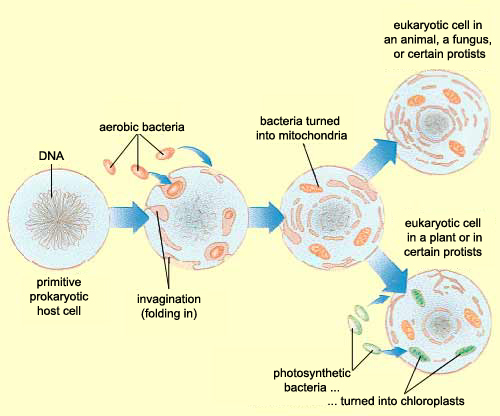
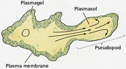
    Zhang et al, instead of assuming that some type of  endogenous bacteria was being released following injury, came up with an interesting hypothesis: it’s not bacteria which is triggering SIRS, but mitochondria. A first year cell biology student will be able to tell you that mitochondria are the parts of eukaryotic cells (sophisticated cells with nuclei) which are responsible for keeping the cell supplied with energy. A long-standing theory in the life science community (pictured above) is that mitochondria, billions of years ago, were originally bacteria which other, larger bacteria swallowed whole. Over countless rounds of evolution, these smaller bacteria became symbiotic with their “neighbor” and eventually adapted to servicing the larger cell’s energy needs. Despite this evolution, mitochondria have not lost all of their (theorized) bacterial ancestry, and in fact still retain bacteria-like DNA and structures. Zhang et al’s guess was that serious injuries could expose a mitochondria’s hidden bacterial nature to the immune system, and cause the body to trigger SIRS as a response.

    Interesting idea, but how do you prove it? The researchers were able to show that 15 major trauma patients with no open wounds or injuries to the gut had thousands of times more mitochondrial DNA  in their bloodstream than non-trauma victims. The researchers were then able to show that this mitochondrial DNA was capable of activating polymorphonuclear neutrophils, some of the body’s key “soldier” cells responsible for causing SIRS.

    Source: Figure 3, Zhang et al.

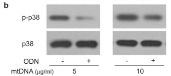
    The figure above shows the result of an experiments illustrating this effect looking at the levels of a protein called p38 MAPK which gets chemically modified into “p-p38” when neutrophils are activated. As you can see in the p-p38 row, adding more mitochondrial DNA (mtDNA, “-” columns) to a sample of neutrophils increases levels of p-p38 (bigger, darker splotch), but adding special DNA which blocks the neutrophil’s mtDNA “detectors” (ODN, “+” columns) seems to lower it again. Comparing this with the control p38 row right underneath shows that the increase in p-p38 is likely due to neutrophil activation from the cells detecting mitochondrial DNA, and not just because the sample had more neutrophils/more p38 (as the splotches in the second row are all roughly the same).

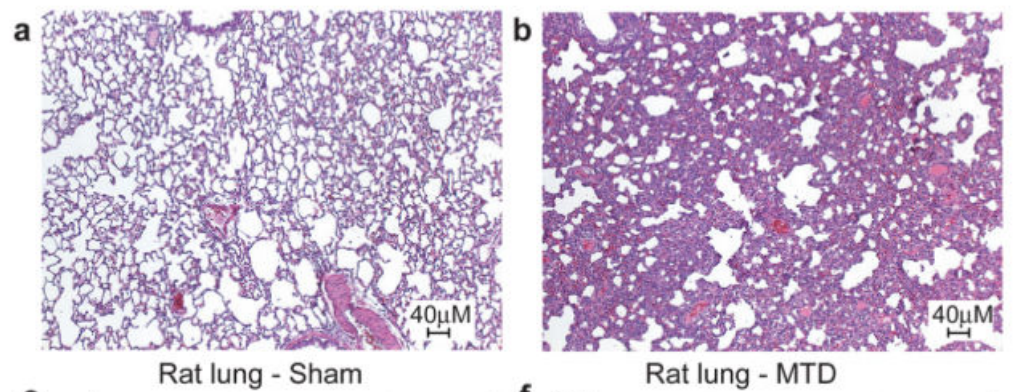
    Cool, but does this mean that mitochondrial DNA actually causes a strong immune response outside of a test tube environment? To test this, the researchers injected mitochondrial DNA into rats and ran a full set of screens on them. While the paper showed numerous charts pointing out how the injected rats had strong immune response across multiple organs, the most striking are the pictures below which show a cross-section of a rat’s lungs comparing rats injected with a buffer solution (panel a, “Sham”) and rats injected with mitochondrial DNA (panel b, MTD). The cross-sections are stained with hematoxylin and eosin which highlight the presence of cells. The darker and “thicker” color on the right shows that there are many more cells in the lungs of rats injected with mitochondrial DNA – most likely from neutrophils and other “soldier cells” which have rushed in looking for bacteria to fight.

    Source: Figure 4, Zhang et al.

    Amazing isn’t it? Not only did they provide part of the solution to the puzzle of injury-mediated SIRS (what they used to call “sterile SIRS”), but lent some support to the endosymbiont hypothesis!

    Paper: Zhang, Qin et al. “Circulating Mitochondrial DAMPs Cause Inflammatory Responses to Injury.” Nature 464, 104-108 (4 March 2010) – doi:10.1038/nature08780

    Check out my other academic paper walkthroughs/summaries

  • Slime Takes a Stroll

    The paper I read for this month brought up an interesting question I’ve always had but never really dug into: how do individual cells find things they can’t “see”? After all, there are lots of microbes out there who can’t always see where their next meal is coming from. How do they go about looking?

    A group of scientists at Princeton University took a stab at the problem by studying the motion of individual slime mold amoeba (from the genus Dictyostelium) and published their findings in the (open access) journal PLoS ONE.

    As one would imagine, if you have no idea where something is, your path to finding it will be somewhat random. What this paper sought to discover is what kind of random motion do amoeboid-like cells use? To those of you without the pleasure of training in biophysics or stochastic processes, that may sound like utter nonsense, but suffice to say physicists and mathematicians have created mathematically precise definitions for different kinds of “random motion”.

    Now, if the idea of different kinds of randomness makes zero sense to you, then the following figure (from Figure 1 in the paper) might be able to help:

    Source: Figure 1, Li et al.

    anel A describes a “traditional” random walk, where each “step” that a random walker takes is completely random (unpredictable and independent of the motion before it). As you can see, the path doesn’t really cover a lot of ground. After all, if you were randomly moving in different directions, you’re just as likely to move to the left as you are to move to the right. The result of this chaos is that you’re likely not to move very far at all (but likely to search a small area very thoroughly). As a result, this sort of randomness is probably not very useful for an amoeba hunting for food, unless for some reason it is counting on food to magically rain down on its lazy butt.

    Panel B and C describe two other kinds of randomness which are better suited to covering more ground. Although the motion described in panel B (the “Levy walk”) looks very different from the “random walk” in Panel A, it is actually very similar on a mathematical/physical level. In fact, the only difference between the “Levy walk” and the “random walk” is that, in a “normal” random walk, the size of each step is constant, whereas the size of each “step” in a “Levy walk” can be different and, sometimes, extremely long. This lets the path taken cover a whole lot more ground.

    A different way of using randomness to cover a lot of ground is shown in Panel C where, instead of taking big steps, the random path actually takes on two different types of motion. In one mode, the steps are exactly like the random walk in Panel A, where the path doesn’t go very far, but “searches” a local area very thoroughly. In another mode, the path bolts in a straight line for a significant distance before settling back into a random walk. This alternation between the different modes defines the “two-state motion” and is another way for randomness to cover more ground than a random walk.

    And what do amoeba use? Panel D gives a glimpse of it. Unlike the nice theoretical paths from Panels A-C rooted around random walks and different size steps or different modes of motion, the researchers found that slime mold amoeba like to zig-zag around a general direction which seems to change randomly over the course of ~10 min. Panel A of Figure 2 (shown below) gives a look at three such random paths taken over 10 hours.

    Source: Figure 2, Li et al.

    The reason for this zig-zagging, or at least the best hypothesis at the time of publication, is that, unlike theoretical particles, amoeba can’t just move in completely random directions with random “step” sizes. They move by “oozing” out pseudopods (picture below), and this physical reality of amoeba motion basically makes the type of motion the researchers discussed more likely and efficient for a cell trying to make its way through uncharted territory.

    Source: 7B Science Online Labs

    The majority of the paper actually covers a lot of the mathematical detail involved in understanding the precise nature of the randomness of amoeboid motion, and is, frankly, an overly-intimidating way to explain what I just described above. In all fairness, that extra detail is more useful and precise in terms of understanding how amoeba move and give a better sense of the underlying biochemistry and biophysics of why they move that way. But what I found most impressive was that the paper took a very basic and straightforward experiment (tracking the motion of single cells) and applied a rigorous mathematical and physical analysis of what they saw to understand the underlying properties.

    The paper was from May 2008 and, according to the PLoS One website, there have been five papers which have cited it (which I have yet to read). But, I’d like to think that the next steps for the researchers involved would be to:

    1. See how much of this type of zig-zag motion applies to other cell types (i.e., white blood cells from our immune system), and why these differences might have emerged (different cell motion mechanisms? the need to have different types of random search strategies?)
    2. Better understand what controls how quickly these cells change direction (and understand if there are drugs that can be used to modulate how our white blood cells find/identify pathogens or how pathogens find food)

    Paper: Li, Liang et al. “Persistent Cell Motion in the Absence of External Signals: a Search Strategy for Eukaryotic Cells.” PLoS ONE3 (5): e2093 (May 2008) – doi:10.1371/journal.pone.0002093

    Check out my other academic paper walkthroughs/summaries